


Amino Acids, Proteins and DNA - Structure of Proteins (A-Level Chemistry)
Structure of Proteins
Peptides, Polypeptides and Proteins
Polypeptides
A polypeptide is a molecule made up of many amino acids, each joined to one another via a peptide bond.
A peptide bond is the amide linkage that forms between the carboxylic acid group of one amino acid and the amine group of another amino acid.
The amide linkage is -CONH- and is shown below:
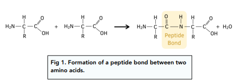
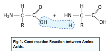
• The reaction between two amino acids is a condensation reaction. This is because a molecule is removed during this process. The OH is removed from the carboxylic acid group of one molecule and the H from the NH₂ group is removed on the other amino acid. These together form a H₂O molecule that is lost.
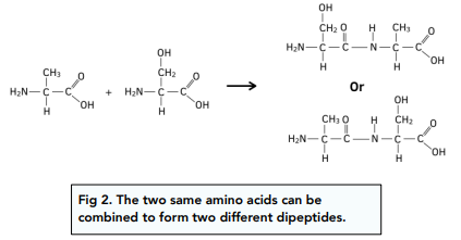
• Two amino acids form a dipeptide. A dipeptide is the molecule formed when two amino acids react together in a condensation reaction. For any two given amino acids, there are two possible combinations of the amino acid in the peptide. This is because both \amino acids contain the same two functional groups.
• More amino acids can be added to a dipeptide to form a polypeptide. The amine and the carboxyl groups will remain intact on either side of the dipeptide so further amino acids can be added in condensation reactions to make a longer polypeptide chain.
Hydrolysis of Polypeptides
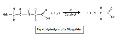
The reverse of the condensation reaction between two amino acids is a hydrolysis reaction between a dipeptide.
The addition of a water molecule can break the peptide bond within a dipeptide will leave two amino acids.
A catalyst of H⁺ ions from highly concentrated HCl is used to hydrolyse the peptide bonds within a polypeptide.
Worked example: A dipeptide is hydrolysed. Identify the structure of the two amino acids it produces.
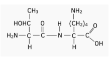
Answer:
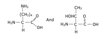
Structure of Proteins
Proteins are made from polypeptide chains. They have a primary structure, secondary structure and a tertiary structure. This results in a complex 3-D shape. They are held together with hydrogen bonds, sulfur-sulfur bonds and weak intermolecular forces.
Primary Structure of Proteins
The primary structure of a protein is the sequence of amino acids in the polypeptide chain, the bonds present in this structure are strong peptide bonds which are strong covalent bonds.
Concentrated HCl and high temperature would be required to overcome these strong bonds within the primary structure of a protein.
An example of primary protein structure is:
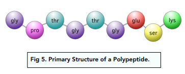
This shows the first three letters of each amino acid in the primary structure.
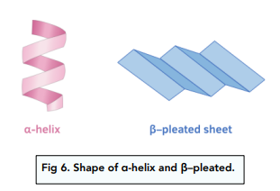
Secondary Structure of Proteins
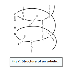
The secondary structure of a protein is either an α-helix or a β–pleated sheet, as shown in the diagram.
The secondary structure of a protein involves hydrogen bonds between amino acids. Different secondary structures have different hydrogen bonding patterns:
• In an α-helix – Hydrogen bonds form between the C=O group of one amino acid and the N-H region of the amino acid 4 places ahead.
This is shown in the diagram.
• In the β–pleated sheet – Hydrogen bonds are also formed between C=O from one peptide link and N-H groups from another peptide link, much further along the chain.
A protein may have several regions which are α-helix and others which are β–pleated sheets.
The secondary structure can be altered by changes in pH or gentle heating. This is because hydrogen bonds are much weaker than covalent bonding.
Tertiary Structure of Proteins
The tertiary structure is a 3D structure formed by the folding of α-helix and/or β–pleated sheets.
The tertiary structure is held together by:
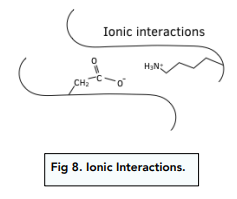
• Ionic interactions – Electrostatic forces of attraction between oppositely charged R groups.
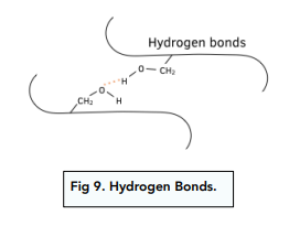
• Hydrogen bonding – Formed between R groups that allow it
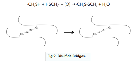
• Disulfide bridges (sulfur-sulfur bonds) – Occur between sulfur atoms within the variable R groups of different amino acids. These S-S covalent bonds are strong and help to fix the tertiary structure of the protein. For example, cysteine is an amino acid with a -CH₂SH group, sulfur-sulfur bonds can form between two of these:
The tertiary structure defines the shape of the protein. This is responsible for the way proteins can act in their biological functions, e.g. in enzymes.
Amino Acid Identification
The first stage in identifying the structure of a protein is to hydrolyse it. This is achieved by reflux with concentrated hydrochloric acid. This breaks the amide bonds and results in a mixture containing all the amino acids.
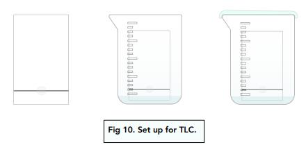
Thin Layer Chromatography
Thin-layer chromatography (TLC) can be used to identify amino acids after hydrolysis of a polypeptide into its constituent amino acids.
Similarly to paper chromatography, a TLC set-up consists of a stationary and mobile phases:
• Stationary phase – The stationary phase is a thin layer of white powder of silica (silicon dioxide) coating a thin, flexible sheet (which would replace the paper in paper chromatography).
• Mobile phase – It is the solvent used in the procedure.
Procedure for Thin-Layer Chromatography
1. Wear gloves. This prevents contamination from hands to the plate.
2. Draw pencil line on the TLC plate. The pencil line should be drawn 1cm above the bottom of the TLC plate.
3. Add the solution on the pencil line. Use a capillary tube to add a tiny drop of each solution being investigated along a pencil line drawn at the bottom of the TLC plate. Then allow the plate to dry.
4. Add the plate to the solvent. Place the plate in a chamber saturated with a suitable solvent. The solvent should be below the pencil line.
5. Put the lid. Immediately replace the lid to get a tight seal.
6. Let the solvent rise up the plate. When it gets to about 1cm from the top, remove the plate and mark the solvent level with a pencil.
7. Dry the plate in a fume cupboard.
8. Spray the plate with a developing agent. Amino acids are colourless. By spraying them with a developing agent like ninhydrin, they will become visible under ultraviolet light.
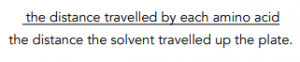
9. Calculate Rf (retention factor) values. A specific amino acid can be identified by comparing its Rf values with a given solvent with a database, using the same solvent. Rf values are calculated as follows:
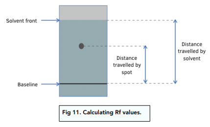
Worked example: A protein is hydrolysed with concentrated acid. Two dipeptides are produced. They are separated by chromatography. The chromatogram is shown below.
Work out the Rf values of both spots
Answer:
A = 0.15
B = 0.55
Amino Acids are the building blocks of proteins, consisting of an amino group (-NH2), a carboxyl group (-COOH), and a side chain (-R) attached to a central carbon atom.
Amino Acids are linked by peptide bonds, formed by the condensation reaction between the amino group of one amino acid and the carboxyl group of another.
A Protein is a large biomolecule made up of one or more polypeptide chains, which are long chains of amino acids linked by peptide bonds.
Primary Structure of Protein is the linear sequence of amino acids in a polypeptide chain.
Secondary Structure of Protein is the local folding of the polypeptide chain, typically into alpha helices or beta sheets, stabilized by hydrogen bonds between the peptide bonds.
Tertiary Structure of Protein is the three-dimensional folding of the entire polypeptide chain, stabilized by a variety of non-covalent interactions such as hydrogen bonds, ionic bonds, hydrophobic interactions, and disulfide bonds.
Quaternary Structure of Protein is the arrangement of multiple polypeptide chains into a single functional unit, stabilized by a variety of non-covalent interactions and sometimes covalent disulfide bonds.
Denaturation of Protein is the loss of the native structure of a protein, usually due to changes in the environment such as high temperature, changes in pH, or exposure to chemicals or detergents.
DNA (Deoxyribonucleic Acid) is a molecule that carries genetic information in cells and is composed of two complementary strands of nucleotides.
The structure of DNA consists of a double helix formed by two complementary strands of nucleotides, each composed of a sugar (deoxyribose), a phosphate group, and a nitrogenous base (adenine, thymine, cytosine, or guanine).
Complementary Base Pairing in DNA refers to the pairing of nitrogenous bases between the two strands of DNA, with adenine (A) always pairing with thymine (T), and cytosine (C) always pairing with guanine (G).
The significance of DNA structure in genetics lies in its ability to carry and transmit genetic information, with the sequence of nucleotides encoding the genetic code that determines the traits and characteristics of an organism. The structure of DNA also enables it to replicate and repair itself, which is essential for the maintenance and transmission of genetic information.
Still got a question? Leave a comment
Leave a comment