


Structure and Function of Blood Vessels (A-level Biology)
Structure and Function of Blood Vessels
The Structure of Blood Vessels
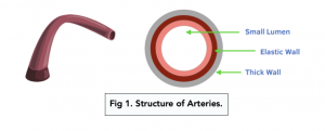
Arteries
- Arteries carry oxygenated blood away from the heart. This filters into arterioles, which lead to the rest of the body.
- Artery walls have thick muscular layers. Thick, muscular walls are needed to withstand the high pressure that arteries are put under
- Thick elastic layers too. Elastic tissue allows the walls to stretch and recoil, to keep in line with the pulsating flow at which the blood travels through.
- Smooth muscle helps blood flow. Smooth muscle lined with smooth endothelium reduces friction and creates less restriction for the blood to move through.
- Overall, the artery wall is very thick. This ensures that the vessel does not burst under pressure.
- There are no valves in arteries. This is because the blood is always under high pressure, so it doesn’t flow backwards. Except in the case of the arteries leaving the heart, which have valves.
Arterioles
- Arterioles carry blood from arteries to capillaries.
- Arterioles are very similar to arteries. They are different in that they are smaller in diameter and have a thinner muscle layer and lumen.
- The muscle layer is thicker and elastic layer is thinner than in arteries. The muscle layer is thicker so that the movement of blood into the capillaries can be controlled, and the elastic layer is thinner because blood pressure is lower in the arterioles.
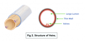
Veins
- Veins transport deoxygenated blood from the body back to the heart.
- The muscle and elastic layers are relatively thin. The muscle layer is thin because constriction isn’t needed to control the flow of blood to the tissues as veins take blood back to the heart. The elastic layer is thinner because the blood is transported slowly and under low pressure, so the veins won’t burst.
- Overall, the wall is not very thick. A thick wall isn’t needed because the pressure within the veins is too low for them to be at risk of bursting.
- Veins have valves. As blood pressure in the veins is so low, the valves ensure that blood doesn’t flow backwards.
- They have a wide lumen. This maximises the volume of blood that is carried to the heart.
- Weak pulse of blood. This indicates that there is very little elastic tissue or smooth muscle as the veins don’t need to stretch or recoil.
Venules
- Larger than capillaries but smaller than veins.
- Blood travels from capillaries into venules, which then branches back into the veins ready to lead towards the heart.
- Venules have thinner walls than arterioles.
- They are porous in order to allow fluid and blood to easily move through their walls.
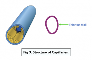
Structure of Capillaries
- Capillaries exchange substances between the blood and body tissues. They are the smallest of the blood vessels.
- Site of metabolic exchange.
- Capillary walls are just one cell thick. They are made up of a single layer of endothelial cells which allows for rapid diffusion of substances.
- There are many capillaries throughout the body and they are highly branched. This means that there is a large surface area for the exchange of substances and that all cells are very close to a capillary.
- Capillaries are extremely narrow. Red blood cells are flattened against the side of the capillary because they are so narrow. This decreases the diffusion distance between the red blood cells and the cells that need oxygen, increasing the rate of diffusion.
- Small spaces are left between the endothelial cells that make up the capillary wall. These spaces allow white blood cells to leave the capillaries and destroy infections in tissues.
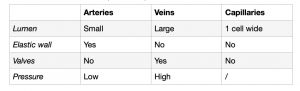
Overview
The table below gives a quick comparison overview:
Pathway of Blood
From the blood starting in your heart, it leaves via the arteries which branch into arterioles. These stem into capillaries where all the nutrients are taken for the cells and the waste products are diffused into the blood. After the capillaries, it moves into venules. Finally, it enters the veins to be carried back to the heart.

The red shows oxygenated blood, blue show deoxygenated blood and purple shows a mixture.

Practical
The practical focuses on being able to identify and draw the structure of arteries and veins.
Equipment:
- Slide with artery section
- Slide with vein section
- Light microscope
- Eyepiece graticule
- Stage micrometer
Precaution:
Be aware of any broken slides as they pose a hazard.
Method:
- Calibration. Calibrate the three objective lens magnifications.
- Place the slide. Carefully, slip the slide under the clips on the microscope stage.
- Lowest power objective lens. Turn the nose piece.
- Coarse adjustment knob. Move the stage closer to the lens. Make sure to not get too close to the lenses.
- Check and adjust. Whilst looking down the microscope, slowly turn the coarse adjustment knob to focus on the sample.
- Fine adjustment knob. Keep turning the fine adjustment knob until the best image is obtained.
- Change lenses. Move to the medium power objective lens and adjust the fine adjustment knob.
- Draw low power plan. This is to show the distribution of tissues, not individual cells. Use the the high power objective lens to help identify the different tissue laters.
- Use the eyepiece graticule. Draw two lines measured in eyepiece units.
- Label. Label your drawing. You should include: endothelium, tunica intima, tunica media, tunica externa and lumen.
- Calculate. Identify the actual size of the low power place and then the magnification of the drawing.
- Repeat. Using the slide with the vein specimen.
Blood vessels are the tubes in the circulatory system that carry blood throughout the body. They include arteries, veins, and capillaries.
The structure of blood vessels includes:
Inner Layer: the inner layer of blood vessels is composed of a smooth, elastic lining called the endothelium.
Middle Layer: the middle layer is made up of smooth muscle and connective tissue, which helps to regulate blood flow.
Outer Layer: the outer layer is composed of connective tissue and provides strength and stability to the blood vessel.
The main functions of blood vessels include:
Transport of Blood: blood vessels transport blood throughout the body, carrying oxygen, nutrients, and waste products to and from the cells.
Regulation of Blood Pressure: blood vessels play a key role in regulating blood pressure by controlling the amount of blood that is flowing through the circulatory system.
Maintenance of Blood Flow: blood vessels help to maintain a constant and adequate flow of blood to all parts of the body, even during changes in physical activity or changes in blood pressure.
Arteries are blood vessels that carry oxygen-rich blood away from the heart to the rest of the body. They have a thick, muscular wall that helps to pump blood through the circulatory system.
Veins are blood vessels that carry oxygen-depleted blood back to the heart. They have thinner walls than arteries and rely on the contraction of the muscles and the pressure of the blood to help return blood to the heart.
Capillaries are the smallest blood vessels in the body and are responsible for the exchange of oxygen, nutrients, and waste products between the blood and the cells. They have very thin walls and allow for the diffusion of materials between the blood and the tissues.
When blood vessels become damaged, they can lead to a range of health problems, including:
Hemorrhage: a condition where there is a loss of blood from the circulatory system.
Thrombosis: a condition where a blood clot forms within a blood vessel, restricting or blocking the flow of blood.
Atherosclerosis: a condition where the walls of the arteries become thick and stiff, reducing the flow of blood to the heart and other organs.
Blood vessel health can be improved through a range of interventions, including:
Exercise: regular physical activity can improve blood flow and reduce the risk of damage to blood vessels.
Healthy diet: a diet rich in fruits, vegetables, whole grains, and lean protein can help to maintain the health of blood vessels.
Avoiding unhealthy habits: avoiding habits such as smoking, excessive alcohol consumption, and a sedentary lifestyle can reduce the risk of damage to blood vessels.
Still got a question? Leave a comment
Leave a comment