


Studying Cells: Confocal Microscopes (A-level Biology)
Studying Cells: Confocal Microscopes
Laser Scanning Confocal Microscopes
- The most common type of confocal microscope is a Laser Scanning Confocal Microscope. Confocal microscopes also use light to illuminate a specimen; however, a Laser Scanning Confocal Microscope (LSCM) floods the specimen with laser (a powerful beam of light) instead of white light.
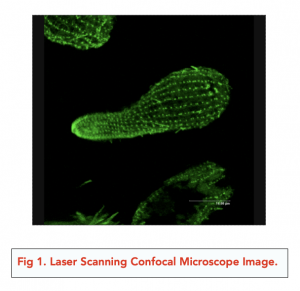
- The specimen is stained with a fluorescent dye. When the laser is shown onto the specimen, this dye fluoresces i.e. it becomes illuminated and gives off its own light. This light that is fluorescing off the specimen is then focused through a very narrow pinhole and onto a detector. The detector may be linked to a computer in order to generate an image, which may be either 2D or 3D.
- LSCM generates high resolution images. The images produced by a LSCM is much clearer (i.e. has a high resolution) compared to images from a normal light microscope. This is because the pinhole blocks off any out-of-focus light.
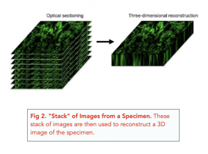
- LSCM is suitable for viewing thick specimens. The laser can scan the specimen multiple times across a plane, and create a “stack” of images. These stacks are then compiled to form a 3D image. This allows us to view the specimen across the entirety of its thickness.
- Limitation: The laser used in older versions of LSCM can damage the cells and tissues in live specimens.
- Limitation: LSCM use more advanced technology than light microscopes, so are much more expensive, especially as they require additional equipment to function such as a detector and a computer. They are also large pieces of equipment and are complicated to set up.
- Limitation: The formation of a “stack” of images can be time consuming as the microscope has to scan the images many times in layers.
Summary of the Different Types of Microscopes
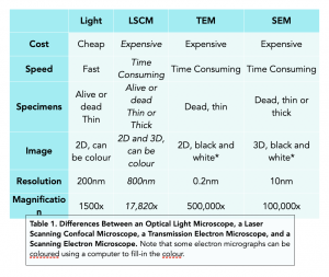
Table 1 summarises the features of the three different microscopes we have looked at.
A confocal microscope is a type of microscope that uses laser light to produce high-resolution images of biological samples. It works by illuminating the sample with a laser, and using a series of lenses and mirrors to focus the light onto a specific plane within the sample. This plane is then scanned, and the light that is reflected back to the microscope is used to create an image of the sample.
The main advantage of using a confocal microscope in cell biology is that it allows researchers to produce highly detailed images of biological samples with minimal damage to the cells. This is because the laser light used in confocal microscopy is tightly focused, and the image is generated by scanning only a specific plane within the sample. This reduces the amount of background noise in the image and improves the overall resolution.
A confocal microscope differs from other types of microscopes in that it uses laser light to produce images of biological samples. This is different from other types of microscopes, such as light microscopes, which use light from a light bulb or LED to illuminate the sample. Confocal microscopes also use a series of lenses and mirrors to focus the laser light onto a specific plane within the sample, which is then scanned to produce an image. This is different from other types of microscopes, such as electron microscopes, which produce images by scanning the sample with a beam of electrons.
There are two main types of confocal microscopes available: single-photon confocal microscopes and multiphoton confocal microscopes. Single-photon confocal microscopes use a single photon of laser light to produce an image, while multiphoton confocal microscopes use multiple photons to produce an image. Both types of confocal microscopes have their own advantages and disadvantages, and the choice between them will depend on the specific needs of the researcher.
Yes, confocal microscopy can be used to study live cells. In fact, one of the major advantages of confocal microscopy is that it can be used to study living cells without causing significant damage to the cells. This is because the laser light used in confocal microscopy is tightly focused, and only a specific plane within the sample is scanned to produce an image.
There are many applications of confocal microscopy in cell biology. For example, it can be used to study the structure and function of cell membranes, to visualize the distribution of specific proteins within cells, and to study the dynamics of cell division and growth. Confocal microscopy is also used to study the organization of cells within tissues, and to visualize the interactions between different cell types in a sample.
Some of the limitations of confocal microscopy in cell biology include the cost of the equipment and the complexity of the imaging process. Confocal microscopy also requires special sample preparation techniques, and can only produce images of samples that are thin enough to allow the laser light to penetrate the sample and reach the focal plane. Finally, some samples may be too thick or too dense to be imaged using confocal microscopy, and may require alternative imaging techniques, such as electron microscopy.
Still got a question? Leave a comment
Leave a comment