


Structure of the Lungs (A-level Biology)
Structure of the Lungs
Gas Exchange
The Route for Gas Exchange
Gas exchange in humans (and most animals) is carried out by a complex organ system, known as the respiratory system.
Though there are many parts to the system, the most commonly thought of is the lungs. The diagram below briefly outlines the process of air entering out body and carbon dioxide leaving:
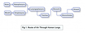
- Breathing in atmospheric air through your nose and mouth travels from your nasopharynx and oropharynx respectively, to your laryngopharynx towards your trachea.
- The trachea then splits into two bronchi (singular. bronchus), each leading into the lungs.
- These branch into bronchioles, smaller tubes ending with small ‘air sacs’ known as alveoli.
- In order to inflate and deflate the lungs to inhale and exhale air, the ribcage, intercostal muscles and diaphragm work alongside each other.
Structures of Gas Exchange
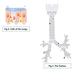
Different types of cells and tissues make up the gaseous exchange system, helping it to work efficiently.
- Goblet cells – these line the airways and secrete mucus in order to trap any microorganisms and dust particles which have been inhaled. It helps prevent them reaching the alveoli.
- Cilia – lining the airway’s cells, cilia work to ‘beat’ the mucus towards the throat to be swallowed, alway from the alveoli. This is efficient at preventing lung infections.
- Elastic fibres – in order to help with the process of breathing out, elastic fibres are in the walls of the trachea, bronchi, bronchioles and alveoli. The lungs inflate and the elastic fibres stretch in order to breathe out. The fibres recoil to push the air out when exhaling.
- Smooth muscle – to be able to control the diameter of the trachea, bronchi and bronchioles, smooth muscle is in their walls. These relax during exercise to be able to expand the tubes, giving less resistance to airflow, letting the air move in and out of the lungs more easily.
- Rings of cartilage – these provide support to the walls of the trachea and bronchi. Whilst strong, it is also flexible in order to strop collapsing when breathing in (and the pressure drops).
Air Entry
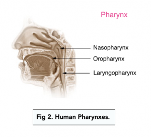
- Air enters the pharyngeal cavities. As you breath in through your mouth and nose, air passes through your oropharynx and nasopharynx respectively. The air then travels into the laryngopharynx.
- The pharyngeal cavities humidify the air. As atmospheric air can be too dry for humans, causing problems, the pharyngeal passages humidify the air.
- The nasal passage is lined with ciliated cells. Ciliated cells in the nasal passage helps to filter out dust, bacteria and occasionally viruses from inspired atmospheric air.
- Mucous membranes line the nasal and oral passages. Mucous membranes in the nasal and oral passages help in humidification of inspired air, but importantly are an immunological barrier and help to trap dust and bacteria.

Trachea
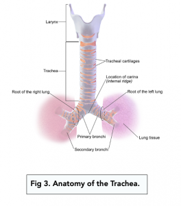
- Inspired air travels from the pharynxes into the trachea. As the trachea is a cartilaginous tube, it is the key airway in most vertebrate organisms. Its main function is to provide a pathway for air entering and leaving the lungs.
- The trachea is adapted to its function:
- Tracheal cartilage – in order to help prevent collapsing, the walls of the trachea are reinforced with specialised cartilage rings called ‘tracheal cartilage’.
- Goblet cells – to be able to trap dust and pathogens, the walls of the trachea are lined with goblet cells which release mucous.
- Ciliated cells – to be able to trap more dust and pathogens, the trachea is also lined with ciliated cells.
Bronchi and Bronchioles
- The bottom of the trachea splits into two bronchi. The bronchi are the two major extensions of the trachea which directly transport the air from the trachea into the lung during inspiration, and air from the lungs into the trachea during expiration.
- The bronchi also have goblet cells and ciliated cells. Similar to the trachea, the bronchi have goblet and ciliated cells to filter out dust and pathogens.
- The bronchi are also supported by cartilaginous rings. Similar to the trachea, the purpose of these rings is to prevent the bronchi from collapsing.
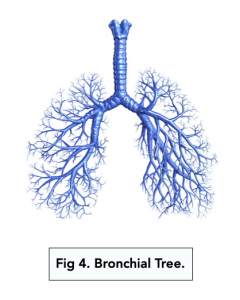
- Each bronchi is split into hundreds of bronchioles. Each bronchi splits into many bronchioles. Bronchioles are simple small, fine extensions of the bronchi which branch off into different regions of the lungs. This is called the bronchial tree.
- Bronchioles have two key types of cells.
-
- The ciliated epithelial cells are there to filter the air of dust and pathogens.
- The smooth muscle cells are responsible for two processes known as bronchodilation and bronchoconstriction.
- The smooth muscles cells of the bronchioles help to regulate their diameter. Bronchioles can dilate or constrict in response to various stimuli from the nervous system.
- Bronchodilation is the widening of the diameter of the bronchioles. Bronchodilation increases the air flow and is caused by the hormone adrenaline. This normally occurs during exercise or during “fight or flight” scenarios.
- Bronchoconstriction is the narrowing of the diameter of the bronchioles. Bronchoconstriction reduces the air flow and is caused by the hormone acetylcholine. This process is not good, and is usually caused by infections, allergies, or excessive stress.
- Bronchodilation and brinchoconstriction happens subconsciously. The two actions are controlled by the sympathetic and parasympathetic nervous systems. In short, this happens subconsciously due to a complex network of hormonal and neurotransmitter signals (don’t worry about this for now).
The Lung
Structure of the Lung
The table below will outline the features associated with different parts of the lung.
|
Part of the lung |
Cartilage |
Smooth muscle |
Elastic fibres |
Goblet cells |
Epithelium |
|
Trachea |
Large (C-shaped) |
Yes |
Yes |
Yes |
Cilia |
|
Bronchi |
Small |
Yes |
Yes |
Yes |
Cilia |
|
Large bronchiole |
None |
Yes |
Yes |
Yes |
Cilia |
|
Medium bronchiole |
None |
Yes |
Yes |
No |
Cilia |
|
Small bronchiole |
None |
No |
Yes |
No |
No Cilia |
|
Alveoli |
None |
No |
Yes |
No |
No Cilia |
The lungs in human beings are a pair of spongy and elastic organs located in the thoracic cavity. They are responsible for exchanging oxygen and carbon dioxide during respiration.
The two main parts of the lungs are the right lung and the left lung.
The anatomy of the lungs is made up of several parts including the bronchi, bronchioles, alveoli, and pleural membranes.
The bronchi are the two main airways that carry air from the trachea into the lungs. They divide into smaller branches, known as bronchioles.
The bronchioles are the small branches of the bronchi and are responsible for carrying air to the alveoli.
The alveoli are small air sacs in the lungs where the exchange of oxygen and carbon dioxide takes place. They are surrounded by capillaries and play a crucial role in respiration.
The pleural membranes are two thin membranes that line the chest cavity and surround each lung. They help to reduce friction during breathing and prevent the lungs from collapsing.
The lungs work in the respiratory system by exchanging oxygen and carbon dioxide through the process of inhalation and exhalation. Air is taken in through the mouth or nose and travels down the trachea into the bronchi and then into the alveoli. Oxygen from the air diffuses into the bloodstream, while carbon dioxide from the bloodstream diffuses into the air in the alveoli.
The structure of the lungs helps with respiration by providing a large surface area for the exchange of oxygen and carbon dioxide. The bronchi, bronchioles, and alveoli all work together to ensure efficient gas exchange. The pleural membranes also help to reduce friction and maintain the shape of the lungs during breathing.
Still got a question? Leave a comment
Leave a comment