


The Ultrastructure of the Sarcomere During Contraction (A-level Biology)
The Ultrastructure of the Sarcomere During Contraction
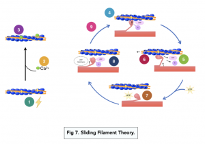
Mechanism of the Sliding Filament Theory
- An action potential crosses the neuromuscular junction. The muscle cell depolarises. The wave of depolarisation passes along the sarcolemma, and spreads down the transverse tubules to the sarcoplasmic reticulum.
- Calcium channels open. Stimulation of the sarcoplasmic reticulum will open calcium channels, releasing stored calcium ions into the sarcoplasm.
- Actin-myosin binding sites are exposed. The calcium ions bind to a protein on tropomyosin, changing the shape of the protein. This moves the tropomyosin and exposes the binding sites.
- Myosin heads bind with the actin-myosin binding sites. The myosin heads form bonds with the actin filaments – these are called actin-myosin cross-bridges.
- ATP is converted into ADP + Pi. The enzyme ATP hydrolase is activated by the influx of calcium ions. The ATP is hydrolysed to provide energy.
- Actin is pulled past the myosin. The energy released from the ATP changes the angle of the myosin head. This allows the actin to slide over the myosin, compacting the sarcomeres and contracting the muscle. This is called power stroke.
- Actin-myosin cross-bridges break. An ATP molecule binds to the myosin head, providing enough energy to break the bond between the myosin head and the actin filament.
- Myosin head reattaches to a different actin-myosin binding site. Hydrolysis of the ATP makes energy available, allowing the myosin to bind further along the length of the actin fibre.
- The cycle is repeated. The myosin head can make and break the cross-bridges up to 100 times a second, so the process is very rapid.

1-1 A-Level Biology Tutoring
Boost your exam scores with personalised tutoring to suit your needs
End of a Contraction
- The cycle can continue as long as calcium ions are present. The sequence ends once all the calcium ions have been pumped back into the sarcoplasmic reticulum by active transport.
- Without the calcium ions, the tropomyosin returns to its original position, blocking the actin-myosin binding sites and allowing the muscle to relax.
Still got a question? Leave a comment
Leave a comment