


The Circulatory System - Heart: Structure and Function (GCSE Biology)
The Circulatory System – Heart
In this section, we will look more in detail at the heart and how it works to supply oxygen-rich blood to the whole body. It is really important for you to understand the structure and function of the heart.
Heart Structure and Function
Chambers of the Heart
The heart has four chambers. Blood is pumped between the following four chambers:
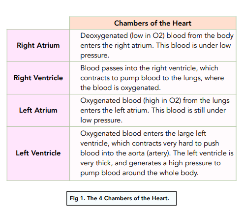
The right and left sides of the heart are divided by a wall called the septum. This ensures that deoxygenated and oxygenated blood don’t mix.
It is the muscular walls of the chambers that contract to generate pressure and pump the blood to its next destination.
Thickness of the Chamber Walls
- Ventricles have thicker walls than the atria – they have to pump blood further at a higher pressure than the atria whereas the atria just have to pump blood to adjacent ventricles.
- The left ventricle has a thicker wall than the right ventricle – the left ventricle has to pump the blood at a higher pressure as the blood will be sent to the entire body whereas the right ventricle only has to send the blood to the lungs which is a much shorter distance so less pressure is needed.
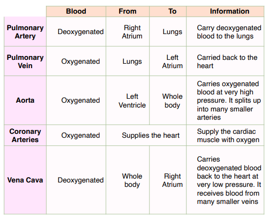
Route of Blood in Vessels of the Heart
The main vessels of the heart mentioned earlier are labelled below alongside the direction of blood-flow:
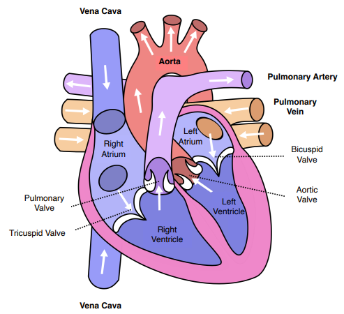

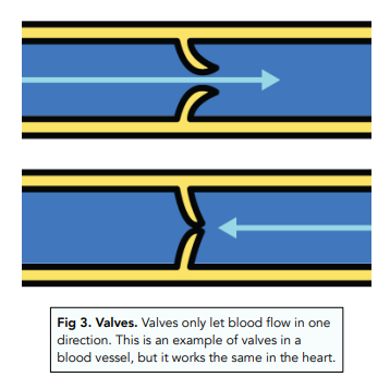
Valves of the Heart
The heart has valves to stop the back flow of blood. Valves are used to keep blood flowing forward. There are four valves in the heart.
- The tricuspid valve: This lies between the right atrium and the right ventricle. It prevents blood flowing back into the right atrium. This is an atrioventricular valve.
- The bicuspid valve: The lies between the left atrium and the left ventricle. It prevents blood flowing back into the left atrium. This is an atrioventricular valve.
- The pulmonary valve: This lies between the right ventricle and the pulmonary artery. It prevents blood flowing back into the right ventricle from the artery. This is a semilunar valve.
- The aortic valve: This lies between the left ventricle and the aorta. It prevents blood flowing back into the left ventricle from the artery. This is a semilunar valve.
Muscle Tissue of the Heart
- The heart contains muscle tissue. Special muscle tissue is present in
the heart. This is called cardiac muscle, and it helps to pump blood
around the body. This tissue is present in the walls of the heart. - Cardiac muscle is myogenic. The muscle is myogenic, which means that it does not tire. Therefore your heart beat continues, keeping you alive!
- The left ventricle has the most cardiac muscle. The wall of left ventricle is the thickest because it has lots of cardiac muscle. This is because the left ventricle is responsible for pumping blood to the whole body, so it needs to contract hard to generate a high blood pressure.
Coronary Arteries
As the heart is made of muscle and it contracts to pump blood to the lungs and around the body, it requires oxygen for aerobic respiration to provide energy for the muscles to contract.
The oxygen is delivered to the heart through coronary arteries which supply the heart with blood.
Activity of the heart
Pacemakers
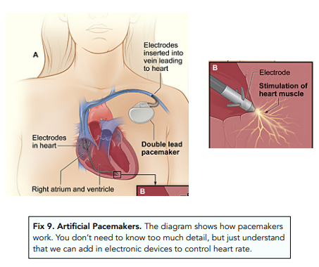
- The rhythm of the heart is controlled by a natural pacemaker. Special cells in the right atrium act as a natural pacemaker for the heart. They keep a resting rhythm in adults of around 70 beats per minute. They do this by sending electrical nerve impulses through the heart muscle, which causes the contraction of the heart, needed to move blood through the heart and into the pulmonary artery and aorta.
- Artificial pacemakers can be used to treat conditions. In some conditions, the heart’s rhythm can be become irregular. Electrical devices called artificial pacemakers can be inserted into the body, which then act to return the heart back to its normal rhythm.
Monitoring Heart Activity
- Using an Electrocardiogram (ECG) – looks at the electrical activity of the heart which is what controls the heart’s activity. Sensors attached to the body are coupled with a machine that gives an ECG reading. Normal and abnormal activity can be identified.
- Using a stethoscope – can listen to the ‘lub-dub’ sounds of the heart made by the valves opening and closing. This can help identify any abnormal sounds.
- Measuring pulse rate – measure how quickly your heart is pumping blood around your body. This can be measured by placing two fingers on the inside of your wrist or at the side of your neck.
Effects of Physical Activity on the Heart
Physical activity will affect your heart rate.
- When you exercise, your body needs more oxygen and produces more carbon dioxide
- Receptors in two arteries detects this increase in carbon dioxide in the blood and send signals to your brain
- The brain signals to the heart to increase its rate and force of contraction
- This sends more oxygen to your muscles You can investigate this yourself by measuring your pulse rate in a
minute before and after exercising. You can even change the intensity of the exercise to explore the effect further
You can investigate this yourself by measuring your pulse rate in a minute before and after exercising. You can even change the intensity of the exercise to explore the effect further.
Effects of Adrenaline on the Heart
Adrenaline is a hormone released by the adrenal glands when an organism feels fright. This travels to the heart via the blood and binds to receptors in the heart telling it to increase its rate and force of contraction.
This is so that more oxygen is delivered to the tissues around the body to prepare it for action (E.g. if it has to run away).
The Circulatory System – Heart: Structure and Function
FAQs
The circulatory system is the body system that is responsible for circulating blood, oxygen, and other essential substances throughout the body.
The heart is a muscular organ located in the chest that plays a crucial role in the circulatory system. It is composed of several layers of tissue and is divided into four chambers: the right atrium, right ventricle, left atrium, and left ventricle. Its primary function is to pump blood throughout the body, delivering oxygen and nutrients to the cells and tissues and removing waste products like carbon dioxide.
The right atrium receives deoxygenated blood from the body and pumps it into the right ventricle. The right ventricle then pumps the blood to the lungs, where it is oxygenated.
The left atrium receives oxygenated blood from the lungs and pumps it into the left ventricle. The left ventricle then pumps the blood out to the rest of the body.
The major blood vessels of the circulatory system include the aorta, vena cava, pulmonary artery, and pulmonary vein.
The aorta is the largest artery in the body and is responsible for carrying oxygenated blood from the left ventricle to the rest of the body.
The vena cava is a large vein that carries deoxygenated blood from the body back to the heart, specifically the right atrium.
The heart regulates the flow of blood in the circulatory system through the contraction and relaxation of its chambers. The coordinated contraction of the atria and ventricles creates the pressure needed to pump blood through the circulatory system.
The heart valves play a crucial role in the proper functioning of the heart by ensuring that blood flows in the correct direction. The heart has four valves: the tricuspid valve, the pulmonary valve, the mitral valve, and the aortic valve.
The tricuspid valve is located between the right atrium and right ventricle, while the pulmonary valve is located between the right ventricle and pulmonary artery. The mitral valve is located between the left atrium and left ventricle, and the aortic valve is located between the left ventricle and the aorta.
When the heart contracts, the valves open to allow blood to flow through. When the heart relaxes, the valves close to prevent blood from flowing backward. This ensures that blood flows in only one direction through the heart and into the circulatory system.
The tricuspid and mitral valves are known as the atrioventricular (AV) valves, as they separate the atria from the ventricles. The pulmonary and aortic valves are known as the semilunar valves, as they have crescent-shaped leaflets that open and close to regulate blood flow.
If the heart valves do not function properly, it can lead to conditions such as valve stenosis (narrowing) or valve regurgitation (leakage), which can cause symptoms such as shortness of breath, chest pain, and fatigue. In some cases, surgical repair or replacement of a valve may be necessary to restore proper function.
Still got a question? Leave a comment
Leave a comment