The Brain - Electrical Stimulation and Scans (GCSE Biology)
The Brain – Electrical Stimulation and Scans
Investigating The Brain
Mapping the Brain
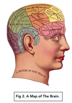
The brain can be investigated in many ways. When research is done into the brain, the aim is to ‘map’ the regions of the brain to particular functions. We mentioned above that the cerebral cortex is divided into over 50 different areas, so for example much of the research has focused on making a map of the cerebral cortex, and working out which are performed which function.

Methods of Investigation
The difficulties of accessing brain tissue inside the skull can be overcome by scanning methods. Key methods of research include:
- MRI scans – MRI scans use magnetic fields and electromagnetic waves in order to investigate activity and the structure of the brain. For example, we can look at the parts of the brains which are most active during different activities and functions (e.g. speaking). MRI scans do not use ionising radiation, so are safer than CT scans.
- CT scans – CT scans use x-rays to investigate the structure of the brain by producing images. They can show damaged structures in the brain which can help you work out why a specific function has been lost following an injury/ disease.
- PET scans – PET scans use radioactive chemicals to highlight activity in the brain. They help you identify any unusual activity in the brain for example, they can show if an area is more active or less active than normal when results are compared with results of a normal brain.
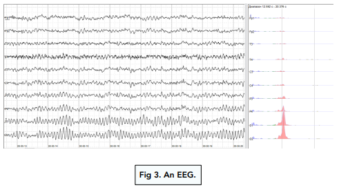
- Electrical stimulation – electrical stimulation is used to map areas of the brain. Certain parts of the brain are stimulated and then the effect is observed. Often the patient is asked what they experienced after stimulation. Electroencephalograms (EEGs) are studied to observe this electrical activity.
- Analysing brain damage – we can look at patients with brain damage to understand the importance of specific parts of the brain. For example, if a patient with poor speech had an abnormal lesion (injury) in a certain part of the cerebral cortex, it is quite likely that this area is involved in speech.

The brain is the complex organ that controls our thoughts, movements, and senses, and is responsible for regulating all of the body’s functions.
Electrical stimulation of the brain is a medical technique that involves using electrical currents to stimulate specific areas of the brain. This can be used to treat conditions such as depression, epilepsy, and chronic pain.
Electrical stimulation of the brain works by using electrodes that are placed on the surface of the scalp or directly on the brain. The electrical currents can increase or decrease the activity of specific areas of the brain, and can modify the signals that are sent between different parts of the brain.
Brain scans are medical imaging techniques that are used to study the structure and function of the brain. They can be used to diagnose and monitor conditions such as strokes, tumors, and degenerative diseases.
The different types of brain scans include computed tomography (CT), magnetic resonance imaging (MRI), positron emission tomography (PET), and functional magnetic resonance imaging (fMRI).
Brain scans work by using different types of energy, such as X-rays, magnetic fields, or radioactive tracers, to create images of the brain. These images can be used to study the structure, function, and blood flow of the brain.
Brain scans can provide information about the size, shape, and activity of different areas of the brain, and can be used to study how the brain responds to different stimuli or tasks. They can also be used to study the effects of drugs, diseases, or injuries on the brain.
Knowledge of electrical stimulation and brain scans can be used to develop new treatments for conditions that affect the brain, such as depression, epilepsy, and traumatic brain injuries. They can also be used to improve our understanding of how the brain works, and how we can maintain brain health as we age.


Still got a question? Leave a comment
Leave a comment